Bacteria - Harmless, helpful or harmful?

Bacteria are . . .
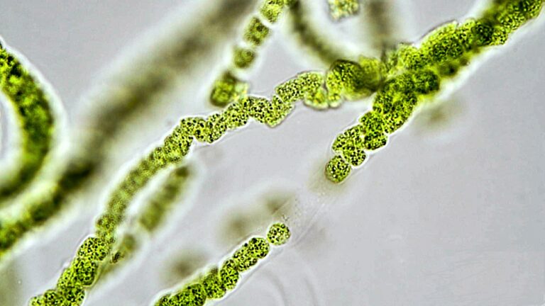
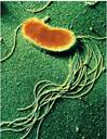
- SINGLE-CELLED organisms found in all types of environments
- Can only be seen through a microscope – measured in micrometers (one millionth of a meter)
- Prokaryotes. Bacteria do not contain a nucleus
- Grouped in families – having the same bacterial ancestors. E.g. Enterobacteriaceae family. A family member is a genus – each genus having developed its own peculiar characteristics. E.g. Escherichia. A genus has different species E.g Escherichia Coli (E. Coli)
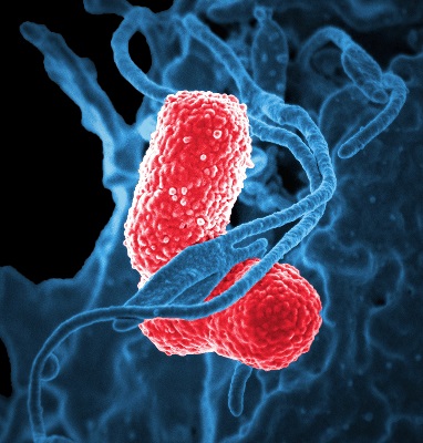
- Different shapes, sizes having many other characteristics
Bacterial purpose / activities
Harmless
Do not affect humans
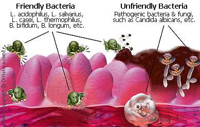
Helpful
Beneficial bacteria produce enzymes that:
- Aid food digestion and production of some vitamins – break down complex food molecules into simpler materials (especially in ruminants). Responsible for production of vitamin K and some B vitamins
- Are involved in production of many foods eaten by humans – milk souring bacteria are used to produce yogurt, cottage cheese, and buttermilk; other bacteria used to produce vinegar and sauerkraut
- Are the principal agents of putrefaction – the anaerobic decomposition of organic materials, especially protein (i.e. contains nitrogen); produces foul odors
- Are the principal agents of decay – the gradual decomposition of organic matter exposed to air by bacteria and fungi.
Harmful (Pathogenic)
Bacteria are potentially pathogenic when:
- Displaced from normal environments in the body – intestinal E. Coli transfer from rectal area into the urinary tractto cause a UTI
- Multiply out of control in an area of the body
- Not part of body’s natural flora and enters body from external source – E.g. Yersinia pestis (responsible for the “Black Plague” in 14th century Europe
Bacteria may produce toxins. Molecules in a cell wall (called adhesins) bind bacteria to the cell and once attached, the bacteria may produce poisonous substances:
- Endotoxins – parts of the cell walls of gram-negative bacteria that are toxic even after the death of the cell. Endotoxins stimulate production of cytokines that can produce widespread vasodilation and shock, and can even cause death
- Exotoxins – enzymes released by bacteria into their host, include hemolysins, leucocidins, coagulases, and fibrinolysins. Exotoxins can also be released in cell lysis (cell death), similarly to endotoxins. These toxins can destroy cells or disrupt normal cellular metabolism. E.g. Clostridium botulinum releases the neurotoxin Botox, that causes botulism. Some toxins are so potent that they can be fatal, since they do not give the immune system time to launch a defense
Pathogenic bacteria cause disease by different mechanisms:
Directly attack tissues (in other life-forms). E.g. Bacterial Leaf Spot caused by Pseudomonas cichorii
Types of bacteria
| Bacterial Type | Characteristics |
|---|---|
| Acetic acid bacteria | Mostly rod-shaped, gram-negative, aerobic; highly tolerant of acidic conditions; generate organic acids |
| Actinomycete (mostly) | Rod-shaped or filamentous, GRAM-POSITIVE, aerobic; common in soils; essential to growth of many plants; source of much of original antibiotic production in pharmaceutical industry |
| Coccoid (mostly) | Spherical, sometimes in clusters or strings, GRAM-POSITIVE, aerobic and anaerobic; resistant to drying and high-salt conditions; Staphylococcus species common on human skin, certain strains associated with toxic shock syndrome |
| Coryneform | Rod-shaped, form club or V shapes, GRAM-POSITIVE,aerobic; found in wide variety of habitats, particularly soils; highly resistant to drying; include Arthrobacter, among most common forms of life on earth |
| Endospore-forming (mostly) | Usually rod-shaped, can be GRAM-POSITIVE or gram-negative; have highly adaptable, heat-resistant spores that can go dormant for long periods, possibly thousands of years; include CLOSTRIDIUM and BACILLUS |
| Enteric(mostly) | Rod-shaped, gram-negative, aerobic but can live in certain anaerobic conditions; produce nitrite from nitrate, acids from glucose; include Escherichia coli,Salmonella (over 1000 types), and Shigella |
| Gliding(mostly) | Rod-shaped, gram-negative, mostly aerobic; glide on secreted slimy substances; form colonies, frequently with complex fruiting structures |
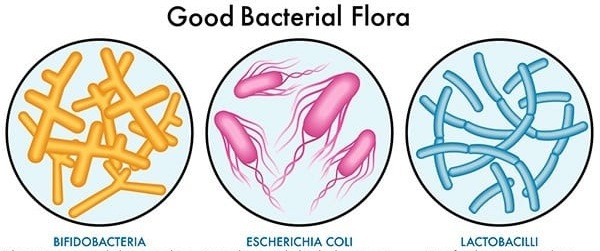
| Lactic Acid Bacteria (LAB) | Rod-shaped or cocci, GRAM-POSITIVE; produce lactic acid through fermentation / break down of food carbohydrates / sugars. Most common LAB in Gi tract are LACTOBACILLUS (essential in dairy product formation), Ligalactobacilli. spp and Bifidobacteria (B. bifidum is prevalent in the large intestine and the intestinal tract of breast-fed infants). Streptococcus (common in humans) Notably S. Mucans is involved in dental plaque and dental caries. LAB are acid-tolerant, CAT-negative, non-sporulating and usually non-motile. |
| Mycobacterium | Pleomorphic, spherical or rod-shaped, frequently branching, no gram stain, aerobic; commonly form yellow pigments; include Mycobacterium tuberculosis, cause of tuberculosis |
| Mycoplasma | Spherical, commonly forming branching chains, no gram stain, aerobic but can live in certain anaerobic conditions; without cell walls yet structurally resistant to lysis; among smallest of bacteria; named for superficial resemblance to fungal hyphae (myco- means ‘fungus’) |
| Nitrogen-fixing | Rod-shaped, gram-negative, aerobic; convert atmospheric nitrogen gas to ammonium in soil; include Azotobacter, a common genus |
| Propionic acid | Rod-shaped, pleomorphic, GRAM-POSITIVE, anaerobic; ferment lactic acid; fermentation produces holes in Swiss cheese from the production of carbon dioxide |
| Pseudomonad | Rod-shaped (straight or curved) with polar flagella, gram-negative, aerobic; can use up to 100 different compounds for carbon and energy |
| Rickettsia | Spherical or rod-shaped, gram-negative, aerobic; cause Rocky Mountain spotted fever and typhus; closely related to Agrobacterium, a common gall-causing plant bacterium |
| Sheathed | Filamentous, gram-negative,aerobic; ‘swarmer’ (colonizing) cells form and break out of a sheath; sometimes coated with metals from environment |
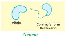
| Spirillum (mostly) | Spiral-shaped, gram-negative,aerobic; include Bdellovibrio, predatory on other bacteria |
| Spirochete (mostly) | Spiral-shaped, gram-negative, mostly anaerobic; common in moist environments, from mammalian gums to coastal mudflats; complex internal structures convey rapid movement; include Treponema pallidum (cause of syphilis) |
| Sulfate- and Sulfur-reducing | Commonly rod-shaped, mostly gram-negative, anaerobic; include Desulfovibrio, ecologically important in marshes |
| Sulfur- and iron-oxidizing | Commonly rod-shaped, frequently with polar flagella, gram-negative,mostly anaerobic; most live in neutral (nonacidic) environment |
| Vibrio (mostly) | Rod- or comma-shaped, gram-negative,aerobic; commonly with a single flagellum; include Vibrio cholerae (cause of cholera), and luminescent forms symbiotic with deep-water fishes and squids |
Reference: Bacteria.Dr. Sayeed Ahmad D. I. Hom. (London)
GRAM POSITIVE or Gram Negative?
The bacterial cell envelope
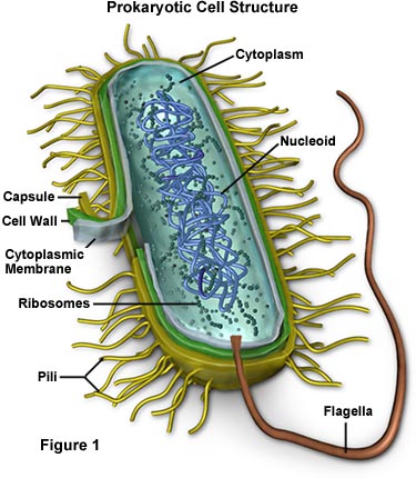
The Cell Envelope comprises the cytoplasmic membrane, cell wall plus an outer membrane, called a capsule (if present):
- Capsule (Only some species of bacteria) – Extra protective – outer capsule composed of polysaccharides (complex carbohydrates). Capsules play a number of roles, but the most important are:
- To keep the bacterium from drying out
- To protect it from phagocytosis (engulfing) by larger microorganisms – a major virulence factor (ability to cause disease) in the major disease-causing bacteria, such as Escherichia coli and Streptococcus pneumoniae. Non-encapsulated mutants of these organisms are avirulent ( i.e. don’t cause disease).
- Cell Wall – Each bacterium is enclosed by a rigid cell wall composed of peptidoglycan, a protein-sugar (polysaccharide) molecule.
- Gives the cell its shape and surrounds the cytoplasmic membrane – protecting it from the environment.
- Helps to anchor appendages like the pili (tiny whiskers used to exchange genetic material) and flagella (whip-like flagellae used to propel bacterium in water). These originate in the cytoplasm membrane and protrude through the wall to the outside.
- Wall strength keeps cell from bursting. The primary function of the wall is to protect the cell from internal pressure caused by much higher concentrations of proteins and other molecules inside the cell compared to outside.
- Unique to bacteria is the presence of peptidoglycan. Located immediately outside the cytoplasmic membrane;
- Cell wall composition varies widely amongst bacteria and is one of the most important factors in bacterial species analysis and differentiation. E.g. Cell wall thickness determines whether bacteria is gram-positive or gram-negative (see below)
- Cytoplasmic membrane. Composed of a phospholipid bilayer, similar to a eukaryotic cell membrane, and similarly acts as a permeability barrier for most molecules and serves as a transport zone for molecules into the cell
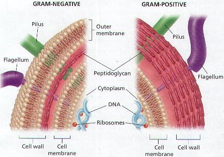
Gram-positive (Thick-walled) or gram-negative (Thin-walled)? In 1884, Danish physician Hans Christian Gram devised a staining and washing technique to differentiate between bacteria with thick cell walls and those with thin walls. When exposed to a “gram stain”, gram-positive bacteria retain the purple color of the stain because the structure of their cell walls traps the dye. In gram-negative bacteria, the cell wall is thin and releases the dye readily when washed with an alcohol or acetone solution.
ACID-FAST bacteria. Won’t hold gram stain, so are difficult to define as gram-positive or gram-negative
| gram-positive | Gram-negative |
| Staphylococcus spp Streptococcus spp Clostridium spp Listeria spp Bacillus Corynbacteria spp | Acinetobacter spp Neisseria Haemo philus Bordetella Helicobacter spp Campylobacter spp Pseudomonas spp Legionella spp Bacteroides Enterobacteriaceae family such as: Escherichia coli Salmonella spp Proteus Klebsiella spp Shigella Citrobacter Serratia Morganella Yersinia Enterobacter |
Peptoglycan layer in the cell wall of Gram-negative bacteria is much thinner than the gram-positive bacteria – comprised of only 20% peptidoglycan
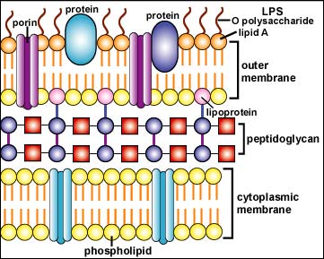
Unique to Gram-negative bacteria is an outer membrane layer – high in lipid content, it is formedby lipopolysaccharide (LPS) layer, lipoproteins, proteins, aphospholipid layer and porins:
- Periplasmic space – separates cell plasma membrane from the peptidoglycan layer (contains proteins which destroy potentially dangerous foreign matter present in this space).
- Phospholipid layer – located in inner layer of outer membrane attached by lipoproteins to the exterior peptidoglycan layer;
Many Gram negative bacteria are pathogenic associated with its endotoxin layer
E.g. The Black Plague wiped out a third of the population of Europe – – It was caused by the tiny G- rod, Yersinia pestis
- PS layer (also known as endotoxin) – this outer layer of the outer membrane is comprised of lipopolysaccharides; their lipid portion is embedded in the outer membrane and is called Lipid A, a toxic substance responsible for most pathogenicity of G- bacteria. Also contributing to toxicity are polysaccharides, called O polysaccharides, extending outward from the bacterial surface.
The highly charged lipopolysaccharides give the G- cell wall an overall negative charge
The center segment, called the core polysaccharide, contains sugars which are highly phosphorylated; it’s these phosphate groups which contribute the negative charge.
- Porins -as a phospholipid bilayer, the lipid part of the outer membrane is impermeable to charged molecules. Channels, called porins, span the outer membrane and allow passive transport of solutes, manyions, sugars and amino acids across the outer membrane into and out of the bacterial cytoplasm
Bacterial shapes and sizes
Size
Average bacillus is 1 μm in diameter by 4 μm in length. They range in size from less than 0.5 to 1.0 μm in diameter to 10 to 20 μm in length for some of the spirilla. For perspective 1,000,000 bacterium placed end-to-end could measure ~ 2 inches.
Shape (3 or 4 main bacterial forms)
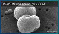
1. Spherical or ovoid (•cocci)
- Coccus (single cells)
- Diplococci (in pairs)
- Staphylococci (clusters)
- Streptococci (chains)
- Sarcinae (cubical groups)
- Tetrad (Group of 4)

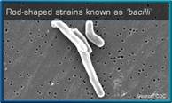
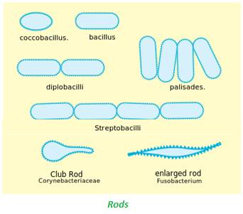
2. Rod-shaped (_bacilli)
- Coccobacilli (more oval)
- Bacillus (single cells)
- Pallisades
- Streptobacilli (forming a chain)
- Diplobacilli (in pairs)
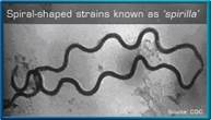
3. Spiral (spirochaete)
- Spirilla (rigid)
- Spirochetes (flexible)
4. Comma (৲ vibro)
- Vibrios (curved, comma).
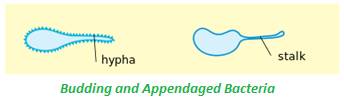
5.Other
Bacterial motility
- Cocci cannot move
- Most bacilli, spiral and comma forms can move independently. Utilizing one or more flagella (slender whiplike appendages used as propellers).
Bacterial growth
acterial growth refers to their reproduction and increase in numbers. Rather than the growth of a single bacterium, which generally doesn’t grow much in size.
The usual method of bacterial reproduction is by binary fission. A process by which a single cell divides to produce two new cells (takes 15 minutes to 16 hours, depending on bacterial type). Alternatively, some bacteria exchange genetic material with members of the same or different species by using their pili (tiny whiskers)
- Reproductive rate affected by changes in temperature, nutrition, and pH
- n an unfavorable environment, some bacilli form self-protective SPORES, called endospores. Their genetic material is condensed and surrounded by a thick wall. Spores are highly resistant to extreme heat, radiation, drying, and chemical agents, such as disinfectants. The spores germinate (revertto growing cells) on return to a favorable environment with nutrients
- Endospore-forming bacteria produce some of the most potent toxins known
- The most clinically significant endospore-forming bacteria. Include Bacillus and Clostridium genera.
Bacterial requirements
Nutritional requirements
- Nutritional requirements of bacteria are much like ours – including sugars, amino acids, vitamins and minerals.
- Bacteria are 80-90% water – too much water flowing into or out of the cell can kill it
- Most bacteria require organic material as food – parasites feed on living organisms, saprophytes feed on non-living organic material
- Some bacteria obtain their energy from inorganic substances (e.g. many soil bacteria) – called autotrophic (self-nourishing).
Temperature requirements
- Most bacteria thrive around human body temperature ( 97°- 99°F) – Although some prefer cold (even freezing) temperatures, others need very hot temperatures (even up to 660°F at cracks in the ocean floor). Each has its own narrow range of temperature in which it can survive.
pH requirements
- Most bacteria thrive in a pH range that is slightly more and less than water (pH 6.5-7.5) – but others can live in a pH more acidic than battery acid
Oxygen requirements
Terms used to describe O2 ReQUIREMENTS for Microorganism GROWTH / REPRODUCTION Group Environment Oxygen (O2) Effect Aerobic Anaerobic Obligate Aerobe
Growth
No growth
Required (utilized for aerobic respiration). E.g. BACILLUS; humans are also obligate aerobes
Microaerophile
Growth if level not too high
No growth
Required but at levels below 0.2 atm; E.g. H. Pylori, Lactobacilli, Camphylobacter
Aerotolerant Anaerobe
Growth
Growth
Not required and not utilized. Lactobacillus natural gut flora
Facultative Anaerobe (Facultative Aerobe)
Growth
Growth
Not required for growth but prefer to use oxygen when available (for efficiency).
E.g. E. Coli, Staphylococcus
Obligate Anaerobe
No growth
Growth
Die in presence of oxygen (and other oxidants).
E.g. Clostridium (however, C. tetani, C. botulinum, C. Perfringens, C. difficile can produce endospores, allowing dormancy / safety);
inefficient energy producers; infect areas of body devoid of oxygen; some use nitrogen compounds to obtain energy; most intestinal bacteria are anaerobes
Bacterial defense mechanisms
Endospores
- An endospore is a dormant, tough, non-reproductive structure produced by certain bacteria from the Firmicute phylum.
- Not a true spore (offspring) – but rather a stripped-down, dormant form of the bacteria
- Endospore-forming bacteria produce some of the most potent toxins known
Usually occurs in GRAM-POSITIVE bacteria – most types of bacteria cannot change to the endospore form, but the most significant clinically significant examples that can are the Bacillus and Clostridium genera:
- Hospital-borne infections: Clostridium difficile
- Food contamination: Bacillus cereus, Clostridium botulinum
- Wound infestation: Clostridium perfringens, Clostridium tetani
- Bioterrorism: Bacillus anthracis
Endospore formation
- When a bacterium detects environmental conditions are becoming unfavorable it may start the process of endosporulation – takes about eight hours
- Endospore formation is usually triggered by a lack of nutrients and endospores can survive without nutrients
- In endospore formation, the bacterium divides within its cell wall then one side engulfs the other
- The endospore consists of: the bacterium’s DNA and part of its cytoplasm, surrounded by a very tough outer coating.
Re-activation
- Endospores enable bacteria to lie dormant for extended periods, even centuries – revival of spores millions of years old has been claimed
- Upon detecting nutrients, the endospore can convert back to actively growing cells – the most common, initial step in this so-called germination process is the recognition of small molecule germinants by germination receptors in the inner-membrane
Endospores are very resilient
- Endospores are resistant to: ultraviolet radiation, desiccation, high temperature, extreme freezing and chemical disinfectants
- Common anti-bacterial agents (that work by destroying vegetative cell walls) do not affect endospores.
Bacterial defense against oxidative damage
Bacteria need to produce the large protein complex cytochrome c oxidase (also called COMPLEX IV or just oxidase) in their cell membranes to survive in the presence of oxygen. If the bacterium produces oxidase in its membrane it isreferred to as OX+, if not then it is called OX-.
Oxidase is essential for bacteria to produce energy using aerobic respiration. Some bacteria can use the efficient aerobic respiratory mechanisms to produce ATP energy molecule from glucose, with oxidase being the last (terminal) enzyme in the respiratory electron transport chain (ETC) required to “harvest” electrons from glucose. (Oxidase catalyzes oxygen’s oxidation of the last molecule in the electron transport chain (ETC) to water – By reducing 1 molecule of oxygen to 2 molecules of water, oxidase translocates 4 protons across the membrane to establish a transmembrane potential difference used by ATP synthase to make ATP energy molecule)
| Oxidase Negative (Ox-) | Oxidase Positive (Ox+) |
|---|---|
| Enterobacteriae (E.g. Escherichia, Shigella, Salmonella, Proteus) | Pseudomonas, Vibrio, Neisseria, Campylobacter (E.g. C. jejuni), Helicobacter (E.g. H. Pylori), Legionella |
Oxidase-positive (OX+) or Oxidase-negative (OX-) ? – an oxidase test determines whether a bacterium produces cytochrome c oxidase and thus can use respiratory mechanism to efficiently produce energy from glucose using oxygen. Bacteria are OX+ if they contain cytochrome c oxidase. OX- bacteria cannot use aerobic respiratory metabolism and are unable to live in the presence of oxygen.
Some bacteria have protective mechanisms against the effects of oxygen. A bacterium’s response to oxygen (or other oxidizers) depends on the presence and distribution of certain enzymes to protect them from damage by oxidation (removal of electrons) – these enzymes react with and neutralize potentially damaging ROS (E.g. Hydrogen peroxide (H2O2), Superoxide Radical (O2.-)) generated by (a) bacterial cells in the presence of oxygen and other oxidizers (E.g. ozone, chlorine, chlorine dioxide) or (b) aerobic cells due to the incomplete reduction of oxygen
SOD, CAT and GPx detoxify oxygen radicals that are inevitably generated by living systems in the presence of O2. The distribution of these enzymes in cells determines their ability to exist in the presence of O2
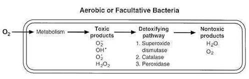
Aerobic or facultative anaerobic bacteria contain enzymes which can detoxify oxygen radicals
Aerobic or facultative anaerobicbacteria contain SOD, CAT or GPx, which can detoxify oxygen radicals produced in the presence of oxygen – converting them to harmless oxygen and water. Obligate anaerobes usually lack or have low levels of SOD, CAT and/or GPx enzymes to remove toxic by-products restricting them to an oxygen-free environment. Obligate anaerobes meet their demise when exposed to oxygen or other oxidizers, being unable to fend them off as the oxidants inactivate their other bacterial enzyme systems.
Superoxide dismutase (SOD)
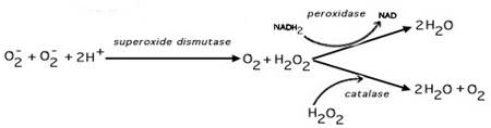
Superoxide dismutase (SOD) enzyme prevents accumulation of potentially lethal superoxide(O2.-) in aerobes and aerotolerant anaerobes. All organisms capable of living in the presence of O2 necessarily contain SOD (regardless of whether they utilize oxygen for their metabolism)
2H++ 2 O2– – –> H2O2 +O2
Catalase (CAT)
Nearly all organisms contain CAT enzymes which catalyze the decomposition of hydrogen peroxide (H2O2) to water
2 H2O2– –> 2 H2O + O2
Catalase-positive pathogenic bacteria make catalase to deactivate peroxide radicals. Thus allowing these bacteria to survive unharmed within the host. Srinivasa Rao PS, Yamada Y, Leung KY (September 2003). “A major catalase (KatB) that is required for resistance to H2O2 and phagocyte-mediated killing in Edwardsiella tarda”. Microbiology (Reading, Engl.)149(Pt 9): 2635-2644.
| Catalase-POSITIVE Bacteria | Catalase-NEGATIVE Bacteria |
|---|---|
| Staphylococci Micrococci Listeria Corynebacterium diphtheriae, Burkholderia cepacia Nocardia The Enterobacteriaceae family (Citrobacter , E. coli, , Enterobacter, Klebsiella, , Shigella,, Yersinia, Proteus, Salmonella, Serratia, Pseudomonas) Mycobacterium tuberculosis, Legionella pneumophila, Campylobacter jejuni, | Streptococcus Enterococcus spp |
Certain aerotolerant bacteria (E.g. lactic acid bacteria such as L. Acidophilus) lack CAT – but can still decompose H2O2 by means of GPx enzymes which reduce peroxide to H2O
Glutathione peroxidase (GPx)(Peroxidase)
Reduces peroxide to H2O utilizing the body’s major in-house antioxidant glutathione
2GSH (Glutathione) + H2O2→GS-SG (Glutathione Disulfide) + 2H2O
| Distribution of SOD, CAT and GPx in prokaryotes with different O2 tolerances | |||
|---|---|---|---|
| Group | Superoxide dismutase (SOD) | Catalase(CAT) | Glutathione Peroxidase(GPx) |
Obligate aerobes and most facultative anaerobes (e.g. Enterics) | + | + | |
Most aerotolerant anaerobes (e.g. Streptococcus pyogenes) | + | – | + |
Obligate anaerobes (e.g. Clostridia, Methanogens (archaea), Bacteroides) | |||
SPECIFIC BACTERIA in disease and microbiome
| Stain (color coded) | Shape | O2 Requirement (color coded) | Other |
|---|---|---|---|
| GRAM POSITIVE g-negative Gram Negative Acid Fast (AF) | • Cocci _ Rod ~ Spiral ৲ Comma | Obligate Aerobe Microaerophile Aerotolerant Anaerobe Facultative Anaerobe Obligate Anaerobe | Motile / nonmotile CAT+ / CAT- Catalase OX+ / OX- Oxidase LAB Lactic Acid Bacteria Encapsulated Endospores |
| Clinically Most Significant Bacteria in the Body | |
|---|---|
| Gram-Negative Bacteria | |
| Bacterial Family Name Genus Species | Associated diseases |
| Gram-Negative Cocci • | |
| Neisseriaceae | g-negative • motile |
| Neisseria | |
| N. gonorrhoeae | Gonorrhea |
| N. meningitidis | Meninigitis |
| Gram-Negative Rods – | |
| Pseudomonadaceae | g-negative _ motile |
| Pseudonomas | |
| P.aeruginosa | Major nosocomial infection hits immunocompromised, IV-lines); UTIs, sepsis, pneumonia, pharyngitis |
| Moraxellaceae | g-negative _ NON-Motile Oxidase-negative |
| Acinetobacter | |
| A. baumannnii | Nosocomial skin and wound infections, pneumonia, meningitis |
| A. iwoff | Meningitis |
| Alcaligenaceae | g-negative_ |
| Bordetella | |
| B. Pertussis nm | Whooping cough |
| B. parapertussis | Mild pharyngitis |
| B. bronchiseptica M | Pneumonia, otitis media |
| Brucellaceae | |
| Brucella | g-negative_ Non-motile |
| B. abortus (from cows) | Bacterial blood infiltration (bacteremia): Brucellosis, Malta fever; |
| B. suis (from pigs) | |
| B. melitensis (from goats) | |
| B. canis (from dogs) | |
| Legionellaceae | g-negative _ |
| Legionella | |
| L. pneumophila | Legionnaires’disease,Pontiac fever, pneumonia |
| L. micdadei | Pneumonia |
| Pseudomonadaceae | g-negative _ Motile |
| Pseudonomas | Major nosocomial infection hits immunocompromised, IV-lines); |
| P. aeruginosa | UTIs, sepsis, pneumonia, pharyngitis |
| Moraxellaceae | g-negative _ Non-motile OX-negative |
| Acinetobacter | |
| A. baumannnii | Nosocomial skin and wound infections, pneumonia, meningitis |
| A. iwoff | Meningitis |
| Pasteurellacae | g-negative _ Non-motile OX-positive CAT positive |
| Haemophilus | |
| H. Influenzae types a-f | Meningitis (Hib) |
| H. influenzae (NTHi / non-typeable) | Otitis media, sinusitis, chronic bronchitis, pneumonia |
| H. aegyptius | Pinkeye (conjuctivitis) |
| H. ducreyi | Venereal disease (Chancroid) |
| Enterobacteriaceae | g-negative _ motilE OX-negative CAT positive and negative |
| Escherichia | |
| E. Coli | UTIs, pneumonia, meningitis, traveler’s diarrhea, peritonitis,pyelonephritis |
| Shigella | Non-motile |
| S. boydii. S. dysenteriae S. flexneri S. sonnei | Dysentry (Diarrhea + fever) |
| Edwardsiella | |
| E.tarda | Gastroenteritis;wound infections |
| Salmonella | g-negative _ |
| 2200 S. species | Bacterial food poisoning |
| S. typhi | Typhoid |
| Citrobacter | |
| C. freundii | Diarrhea? |
| C. diversus | Meningitis in newborns? |
| Klebsiella | |
K. pneumoniae | UTIs |
| Enterobacter | |
| E. aerogenes (F) | UTIs |
| E. cloacae | UTIs |
| Serratia | |
| S. marcescens | Utis,, wound infections |
| Proteus | |
P. mirabilis, P. vulgaris | UTIs, wound infections,Hospital acquired infections |
| Morganella | |
| M. morganii | UTIs, wound infections,diarrhea |
| Yersinia | |
| Y. enterocolitica | Intestinal inflammation / pain(via enterotoxin release) |
| Y. Pestis | The Plague / Black Death in the 14th century; Bubonic, pneumonic, and septicemic plagues |
| Gram Negative Curved Rod / Comma ৲ | |
| Vibrionaceae | g-negative ৲ motile OX-positive CAT-positive |
| Vibrio | |
| V. cholerae | Cholera |
| Gram Negative Spiral~ | |
| Campylobacteraceae | g-negative ~ motile |
| Campylobacter | |
| C. jejuni | Diarrhea, gastrointestinitis |
| Helicobacteraceae | g-negative ~ (helix) motile OX-positive CAT-positive |
| Helicobacter | |
| H. Pylori | Stomach and duodenal ulcers, Gastric carcinomas, Chronic gastritis (in U.S.) |
| Bacteroidaceae | |
| Bacteroides | motile or NON-motile. Commonly found in human gut flora |
| Spirochaetaceae | g-negative ~ motile |
| Borrelia | |
| B.burgdorferi | Lyme Disease |
| Treponema | |
| T. pallidum | Syphilis, yaws |
| GRAM-POSITIVE Bacteria | |
| Bacterial Family Name Genus Species | Associated diseases |
| GRAM-POSITIVE COCCI• | |
Micrococcaceae | G-POSITIVE • NON-motile OX+ CAT+ |
| Micrococcus | |
M. luteus, M. roseus, M. varians | Harmless skin contaminant, |
| Staphylococcus | Soft tissue infections, TSS; |
S. aureus | Scalded skin syndrome, cellulitis, pneumonia, meningitis, boils, arthritis, osteomyelitis |
| Streptococcus | LAB |
| S.pyogenes | 90% of pharyngitis (“strep throat”) |
| S.pneumonia | Pus-producing; Pneumonia, meningitis, otitis media |
| S.mutans, s. mitis (Viridans group) | Large percentage of tooth decay |
| ENTEROCOCCaceae | G-POSITIVE • |
| Enterococcus | LAB; Species leading cause of nosocomial infections (especially catheterizations, reinfected root canal-treated teeth) e,g, endocarditis, UTIs |
| E.faecalis | nm Naturally inhabits GI tract (90-95%); opportunistic pathogen |
| E.faecIUM | Intestinal bacterium (5-10%) |
| GRAM-POSITIVE RODS _ | |
MYCOBACTERIACEAE | G-POSITIVE / AF _ Non-motile |
| M. tuberculosis | Tuberculosis |
M. Leprosy | Leprosy |
| BACILLACEAE | G-POSITIVE _ CAT+ |
BACILLUS | |
B. AnthraCIS | Non-motile Anthrax |
B. CEreUS | motile Toxin-mediated food poisoning |
| LACTOBACILLACEAE | G-POSITIVE produce lactic acid through |
| Lactobacillus | Commonly used as part of the fermentation process in yoghurt, cider, wine, sauerkraut, pickles, cheese, chocolate, and other fermented foods. Common in GI tract; only LAB producing solely LACTIC ACID (homofermentative). Different strains of the same species do different probiotic “jobs” in the body. |
| L. acidophilus strains | Probiotic |
| L. Bulgaricus | Probiotic; only converts lactose => Lactic Acid |
| L. plantarum strains | Probiotic |
| L. Reuteri strains | Probiotic |
| L. Rhamnosus strains | Probiotic |
L. PARACASEI strains | Probiotic |
| Lacticaseibacillus | Common in GI tract; |
| L. casei, | Probiotic; Related to cheese |
| LigalactoBacillus | Common in GI tract;; Harmless; ferments glucose or lactose => lactic acid (homofermentative); considered oxygen-tolerant |
L. brevis, L. animalis | PProbiotics. |
| LISTERIACEAE | G-POSITIVE _ CAT+ |
| LISTERIA | |
| L. monocytogenes | Food poisoning, septocemia, meningitis in immuno-compromised; listeriosis |
| PROPionibacteriaceae | G-POSITIVE _ |
| PROPionibacterium | Acne, chronic blepharitus |
| P. ACNES | |
| CORYNEBACTERIACEAE | G-POSITIVE _ |
| Corynebacteria | |
| C. Diptheriae | Diptheria (caused by toxin) |
| Actinomycetaceae | G-POSITIVE _ (resemble fungi) Produce spores |
| Actinomyces | |
| A. Israeli | Most common cause of infection in dental procedures and oral abscesses |
| A.Nocardia | Pulmonary disease, cutaneous infections |
| Bifidobacteriaceae | G-POSITIVE _ NON-motile |
| Bifidobacteria | Probiotic. Common in GI tract; LAB |
| B. Bifidum | Most predominat bacteria in infant GI tract. |
| Clostridiaceae | G-POSITIVE _ |
| Clostridium | |
| C. Botulinum, | motile Botulism: from botulinum neurotoxin (Botox), usually food-borne. Honey can contain spores affecting infants <1yr old) |
| C. butyrculum C. barati C argentinense | |
C. Difficile | motile Pseudomembranous colitis; common nosocomial infection |
| C. Tetani | motile Tetanus |
| C. Perfringens | NON-motile Gas gangrene, clostridial necrotizing enteritis |
| PEPTOSTREPTOCOCCUS | |
| P. Magnus | |
| P. anaerobius | Infections of oral cavity, respiratory, female genitourinary and GI tracts, bone and joints; also deep organ abscesses and leg/foot ulcers |
| P. micros | |
References
Taber’s Online Medical Dictionary
http://www.scienceclarified.com/As-Bi/Bacteria.html#ixzz2JqhuVa1t
Wikipedia