Central Nervous System (CNS) - Brain and spinal cord

The spinal cord
Vertebral column
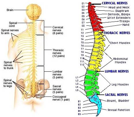
Vertebrae (bones)
- Cervical (7 vertebrae): Located in the neck.
- Thoracic (12 vertebrae): Located in the upper and mid-back.
- Lumbar (5 vertebrae): Located in the lower back.
- Sacral (5 fused vertebrae): Located in the pelvic region.
- Coccygeal (4 fused vertebrae): Forming the coccyx or tailbone.
Posterior longitudinal ligament. Separates invertebral discs from tubal spinal cavity / canal, which houses and protects the nerve-carrying spinal cord, from base of skull to lower back.
Meninges. 3 protective membranes surround spinal cord
Invertebral discs. Providing cushioning and flexibility, a connective, spinal disc, composed of strong elastic tissues, separates each vertebra. When the spine bends or rotates, these spinal discs allow smooth, low-friction movement between adjacent vertebrae. Each disc contains a soft inner substance with a surrounding outer layer, which maintains the structure of the disc.
When the tough outer portion of the disc weakens, it is possible for the gel-like center of the spinal discs to bulge outwards into the spinal canal.
In more severe cases, it can lead to a herniated disc. A herniated disc occurs when a tear in the outer layer of the disc allows the inner substance to leak into the spinal canal.
The brain
The brain exists at birth but continues to develop through your 20’s until it weighs an average of 3 pounds
The brain has several functions
The brain sends, receives and processes signals, that carry information to and from your organs and muscles, your senses (sight, smell, sound, touch and taste), and other areas of your body to relay information and initiate required action regarding such as pain, temperature, heart rate, movement and speech. The brain uses this information to help you can understand and interact with the world around you.
- “Behind-the-scenes, automatic activities E.g. breathing, heart rate, sleep and temperature control
- “Fight-or-flight” stress response
- Memory storage and recall, emotional balance
- Maintaining your body’s organs
- Sensory processing (vision, hearing, smell, touch and taste)
- Movement, balance, coordination
- Speech and language
- Decision-making, prioritizing tasks, emotional control. Handled by the prefrontal-cortex – the last part of the brain to mature in your mid-late 20’s
The brain has two types of cells
(1) Neurons (nerve cells)
Neurons carry information to and fro in the CNS. Using electrical impulses and chemical signals they transmit messages between different parts of the brain, and between the brain and the rest of the nervous system. This allows us to interact with our world and empowers our movement, thoughts, emotions and desires.
The neuron comprises 3 main parts:
- Cell body (soma). Includes nucleus (contains DNA, regulates cell activities), and organelles (e.g. ribosomes, mitochondria, endoplasmic reticulum, and Golgi apparatus).
- The dendrites. Branches protruding from cell body, receive chemical signals (messages) from other neurons, convert them to electrical impulses and transmit them to the cell body.
- The axon. Long extension that conducts impulses from the cell body to the axon terminal, where the signal is passed on to target cells. It is usually surrounded by an insulating myelin sheath that increases conduction speed.
3 types of neurons:
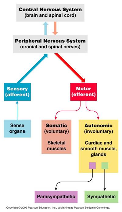
- Sensory (afferent). Activated by stimuli (e.g. light, sound, heat, pressure) outside neuron, they carry sensory information from the peripheral nervous system (PNS), e.g. from the eyes, ears, fingertips to the brain. For example, if you touched a hot pan.
- Motor (efferent). Carry impulses from the brain to the muscles,via the PNS to control our voluntary movements. E.g. The brain tells you to move your hand away from the aforementioned hot pan.
- Interneuron. Most abundant of the 3 types, exclusive to the CNS, they act as a “middleman” between sensory and motor neurons. It also connects to other interneurons, allowing them to communicate with one another.

(2) Glial cells
.Glial cells (also called neuroglial cells) originally named because they hold neurons together (Glia is Greek for glue), but it is now known that they also have an active role in brain signaling. They communicate with neurons and other glial cells via chemical signals.
There are 4 types of glial cells in the CNS: the astrocytes, oligodendrocytes and ependymal cells (collectively known as the macroglia), and the microglia,
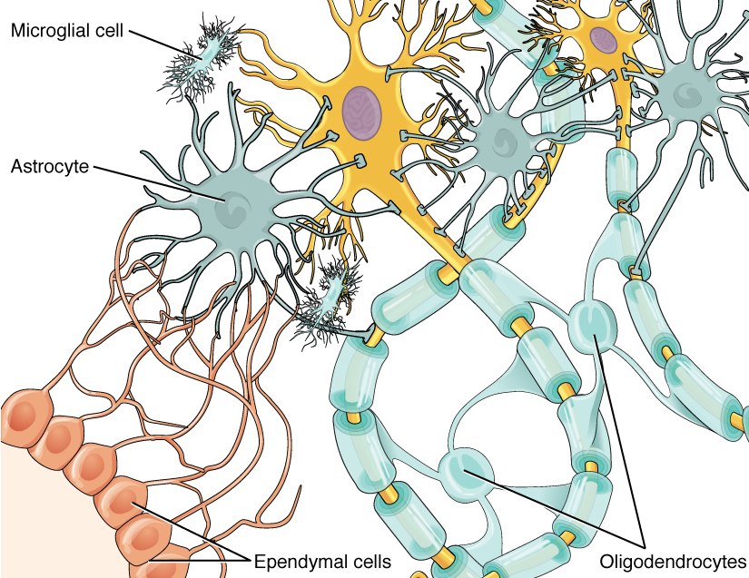
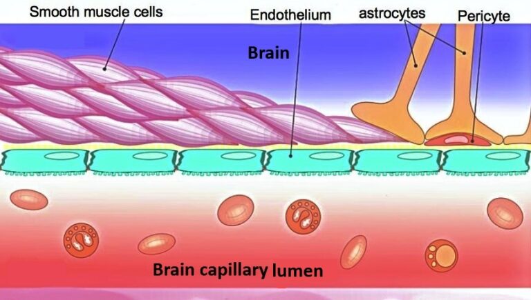
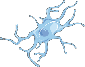
Astrocytes. Named for their star-like shape. their main function is to maintain the integrity of blood brain barrier (BBB). Their “end-feet” at the end of their protrusions are often wrapped around the endothelial cells (ECs) that surround blood vessels to provide the ECs the compounds they need to maintain a healthy BBB.
Astrocytes produce trophic factors, chemicals to promote growth and development of neurons, and signal neurons to continue to live or to maintain specific synapses.
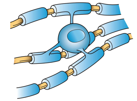
Oligodendrocyte. Only in the CNS, one oligodendrocyte produces enough myelin (a fatty substance) to form a myelin sheath around about 50 nearby segments of neuronal axons, to allow for rapid conduction of electrical impulse signals along the length of their axons.
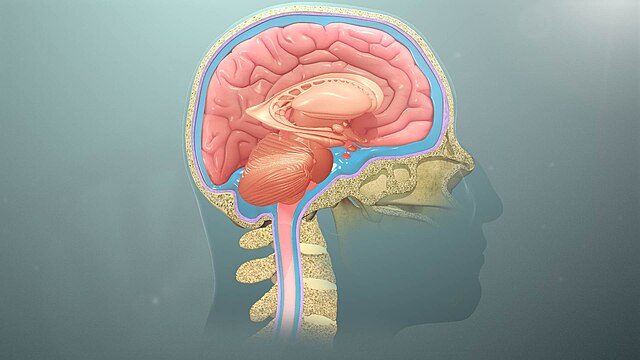
Ependymal cells. Produce over 2 cups of cerebral spinal fluid (CSF) in the body each day. They form a lining inside the spinal cord and cavities within the brain (called ventricles), which are filled with CSF.
Microglia. CNS immune cells protect the brain and spinal cord from damage and disease by identifying and removing infectious agents, material in the synapse, damaged myelin, and dead or damaged cells.
The traditional view that microglia remove protein aggregates has been upended with recent findings that the dense-core amyloid beta (Aβ) plaques, a hallmark of Alzheimer’s disease. are actually promoted and formed as a defensive mechanism by the action of microglia (with surface proteins called TAM receptors), as they sweep wispy Aβ plaque away from neurons, which would otherwise cause their death. After engulfing the Aβ, microglia transfer it to a dense core plaque – a beneficial mechanism since it gets harmful Aβ plaque away from neurons. Some Alzheimer’s Plaques May Be Protective, Not Destructive, 2021 An author of the research study suggested that “when there are fewer dense-core plaques, there seem to be more detrimental effects”, and “we seem to find that dense-core plaques are a bit more benign.” The focus should not be on breaking up the aggregates, but finding out what causes the Aβ plaque. Aβ is also present in Parkinson’s disease (PD) and dementia.
Considered a hallmark of Parkinson’s disease, amyloid fibrils comprised of misfolded α-synuclein (α-syn) proteins, form aggregates, called Lewy bodies. α-syn is a protein abundant in the brain involved in the release of neurotransmitters.
Amyloid plaques block cell-to-cell signaling at synapses, a process essential for storing memories, processing thoughts and emotions, and planning.
The brain has 3 main parts
Cerebrum – Largest part of brain (right and left hemispheres)
- Interprets the senses
- Regulates actions of your conscious thoughts. E.g. speech, memory, behavior, personality, movement, reasoning and judgment
Cerebellum – small semicircle in back of brain
Brainstem – lower part of brain connecting brain to spinal cord
- Maintains involuntary (unconscious) functions. E.g. heart rate, breathing, sleeping / waking cycles, swallowing
A bony structure called your cranium surrounds your brain. Your cranium is part of your skull. Your brain floats in a liquid called cerebrospinal fluid (CSF). All the bones of your skull and CSF protect your brain from injury.
The brain is well-protected from internal and external injury
The skull (cranium) is a bony structure comprising (1) facial bones that support the face and (2) the braincase.
Floating in cushioning cerebral spinal fluid (CSF) inside the braincase the brain is afforded protection against traumatic injury.
The blood-brain-barrier (BBB) provides protection against harmful substances or infectious microbes entering the brain