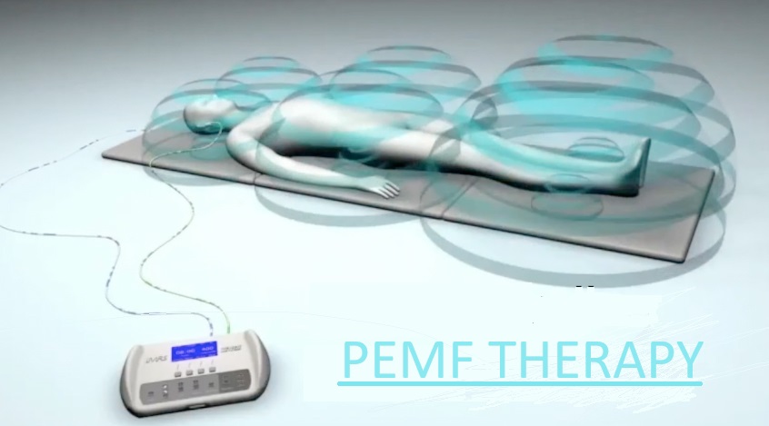
Healing effects of pulsed electromagnetic field (PEMF) therapy in the body
* IMPORTANT * – Re: SOTA instruments Please read this disclaimer
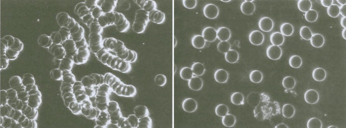
When red blood cells (RBCs) clump together (called the Rouleau effect), oxygen uptake is reduced. PEMF therapy not only separates the RBCs enabling them to better take up oxygen, but also enables them to better fit in the small capillaries to deliver oxygen to cells.
Rapidly changing (Pulsed) electromagnetic fields (EMFs) present several useful therapeutic phenomena when applied to animal / human tissue. Results vary with therapeutic strength, rate of change of magnetic field, and duration of change of field. PEMF therapy upregulates the following inter-related functions, providing the body optimal means to heal itself:
- Normalizes the cellular transmembrane potential (TMP or “cell battery” voltage)
- Decreases pain and inflammation
- Increases blood oxygen levels and improves circulation
- Improves cellular ATP energy production
- Increases vessel-dilatory NITRIC OXIDE production
- Restores immune System Function
- Deactivates micro-organisms
- Promotes tissue repair / healing
- Destroys cancer cells
- Increases cellular pH (Alkalizes)
- Other effects
Normalizes the cellular transmembrane potential (TMP or “cell battery” voltage)
A cell’s transmembrane potential (“cell battery” or cell membrane capacitance) serves 2 purposes.
(a) It provides power for cellular operations (e.g. mitochondrial production of ATP energy, cellular metabolism) and (b) It is used for transmitting signals between different parts of electrically excitable cells, such as neurons (e.g. to transmit a pain message from the brain) and muscle cells (to make a muscle contract).
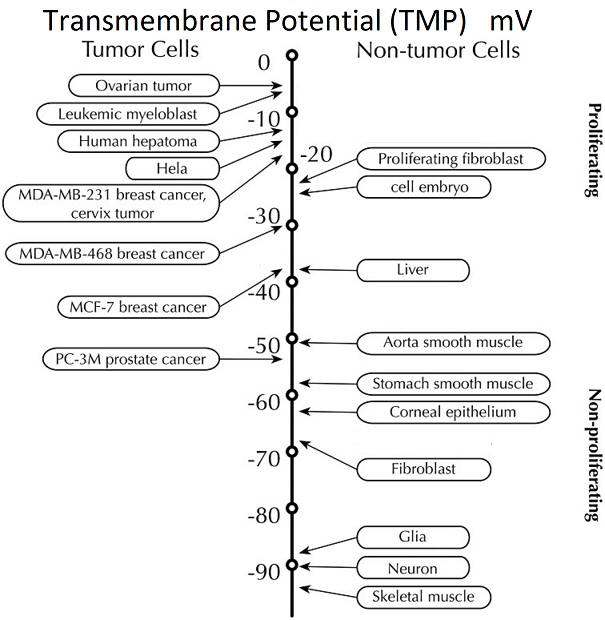
The usual convention when referring to a cell’s transmembrane potential difference (or “cell battery” voltage) is to define it as negative – meaning that the inside of the cell is more negative than the outside. E.g. a TMP of -90mV means a there is a difference of 90mV across the membrane – the “cell battery” voltage is 90 mV.
Much of the success of PEMF therapy lies in its ability to significantly raise the generally lower ““cell battery”” voltage of sick or damaged cells to a normal, healthy range. E.g. Lower than ideal / normal TMP levels can functionally regulate tumorigenesis and promote cancer progression. (Yang & Brackenbury, 2013)
Natural PEMFs of the earth enhance the sodium / potassium (Na++/K++) pumps in every cell membrane to establish a healthy “battery” voltage across the membrane. The cell membrane separates and maintains a balance between the intracellular and extracellular fluid and electrolytes, such as positively charged Na and K, and negatively charged chlorine (Cl) ions. The Na/K pump’s job is to move 3 Na ions OUT of the cell and 2 K ions IN through transmembrane protein channels, to maintain a healthy net positive charge on the outside of the cell membrane. At least 30% of the ATP energy produced by the cell is used to fuel the important Na/K pumps. Cell membranes are capacitors, able to accumulate and store charge (energy) to be given up when needed. Any condition, illness or change in dietary intake that affects the composition of the cell membranes and their associated minerals can affect and alter cellular capacitance.
By establishing the “cell battery” voltage, the Na/K pump has an essential role in all cellular functions: including bringing nutrients into the cell for the mitochondrial production of ATP, cellular metabolism, and removing cellular metabolic waste products and toxins.
Transmembrane potentials ( “cell batteries”) of living cells will not accept a charge greater than in the normal range. Similar to recharging any battery.
Low frequency electromagnetic signals (from the body or external environment) can open and close cell membrane ion-channel gates. This too may affect maintaining the voltage of the “cell battery”, by helping ions cross the cell membrane.
Decreases pain and inflammation
(by restoring normal cellular transmembrane potentials at an accelerated rate)
Pain is your body’s way of alerting you to a problem so that you can address and heal that problem. Ideally, the brain produces endorphins, endogenous morphine or other natural opiates, to control or reduce our perception of pain by interacting with the opiate receptors in the brain. Endorphins are brain neurotransmitters, which transmit electrical signals within the CNS for an analgesic effect.
Addictive, endorphin drugs (such as morphine, Vicodin, Percocet, Oxycontin, heroin and opium) override the body’s natural endorphin production.
Endogenous endorphin production is increased by: exercise, keeping stress levels low (yoga / deep breathing / meditation / massage), REM sleep, laughter, sex and earth-based (low) frequencies, including those generated in PEMF therapy
PAIN AND THE CELL TRANSMEMBRANE POTENTIAL (TMP) CONNECTION
- All cells have a potential difference between their inner and outer membrane. A nerve or muscle cell has a normal resting transmembrane potential (TMP or “cell battery” voltage) of ~70mV and emits few chemical pain signals and inflammatory agents (histamines, NITRIC OXIDE, prostaglandins, etc).
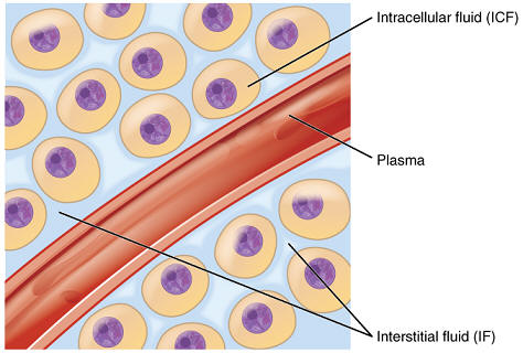
- When cells are damaged, their transmembrane potentials (TMPs) decrease to about 80% of healthy levels. (Ceve, 1990; Malzone). This causes positive sodium ions to enter cells and potassium, and negative trace elements and proteins to exit cells. Damaged cells release chemical signals, such as histamine, which increase capillary blood flow to affected area. Capillaries also pass fluid into the interstitial space between cells, which accumulates to cause interstitial swelling or edema with related pain.
- PEMF therapy reduces edemic swelling / pain outside damaged cells. PEMF therapy applied to inflamed or painful sites increases the transmembrane potential of damaged cells (i.e. recharges their “cell batteries”) and so reduces edema.
- PEMF therapy lowers neural pain transmission. Pain signals are transmitted along nerve cells (neurons) to their pre-synaptic terminals. Triggered by a large decrease in the neuron’s resting “battery” voltage, the neuron releases a chemical transmitter from a synaptic vesicle contained within the membrane to be received by the next adjacent neuron, and so passes along the pain message.
The application of PEMF therapy to inflamed or painful sites causes “battery” voltage of neurons to be raised to a hyper-polarization level of about 90 mV, preventing transmission of a pain signal.
| PEMF THERAPY BLOCKS PAIN SIGNAL TRANSMISSION FROM NEURON TO NEURON |
|---|
Pain signals are transmitted (by the movement of ions) along a nerve cell, and then from that nerve cell to another nerve cell by releasing a chemical neurotransmitter across the synaptic gap between them. The neurotransmitter release is triggered when the voltage across the nerve cell’s membrane at the synaptic gap drops to -30mV. The membrane voltage at the synaptic gap is about 70mV when under “no pain” conditions, but decreases to -30mV as the pain signal approaches. The 100mV (70mV to -30mV) average change of potential voltage is sufficient to trigger the neurotransmitter release and thus transmit the pain signal to the next cell. i.e. the brain eventually receives the pain signal. However — when PEMF therapy to painful areas raises the membrane voltage to about 90mV, the 100mV change lowers the synaptic voltage to only -10mV (90mV – 100mV, which is insufficient to release the neurotransmitter, and therefore blocks the transfer of the pain signal. |
Specific frequencies regulate / balance neurotransmitters in the neuroendocrine system. E.g. Endorphins and DOPAMINE (also associated with pain). For soft tissues, low frequency (natural or applied) electromagnetic fields create currents conducted primarily along the surface of cells, which amplify weak triggers associated with the binding of neurotransmitters (also hormones and antibodies) to their specific cell binding sites (Adey, 1993).
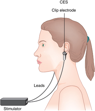
Therapeutic frequencies do not necessarily have to be applied to the head region, since the meridians can carry them throughout the body.
Increases blood oxygen levels / improves circulation
Oxygen is supplied to the body via the respiratory system from the alveoli in the lungs, attached to hemoglobin in red blood cells in the pulmonary capillaries, from where it is carried into the circulatory system. Circulating RBCs deliver their oxygen cargo and other nutrients to cells and also transport cellular waste, such as carbon dioxide, hydrogen peroxide, and toxins, to the elimination organs via the microcirculation of the capillaries. Optimal cellular ATP energy production occurs with a sufficiency of oxygen provided by optimally functioning respiratory and circulatory systems. PEMF therapy improves these via the following effects:
PEMF therapy increases blood flow and oxygen delivery in capillary microcirculation
- Capillaries are the small vessels at the interface where oxygen diffuses into cells and cellular waste is carried away
- PEMF therapy decreases red blood cell (RBC) aggregation by increasing their cellular TMP (“cell battery” voltage). When RBCs clump and stack together (called the Rouleau effect), oxygen uptake is reduced. Additionally, stacked RBCs can not fit in the capillaries to deliver oxygen to cells, since their small lumens require that RBCs travel in single file. When the RBC “cell battery” voltage is high, there is a strong positive charge on the outside of cells, which can repel other cells with a similarly high “cell battery” voltage, thus keeping RBCs apart from each other.
PEMF therapy improves general circulation
- PEMF therapy lowers blood (and lymph) viscosity. This enables blood (and waste-carrying lymph) to flow more easily, promoting lower blood pressure and reduced risk of arterial blockage through areas with atherosclerotic build-up.
- PEMF therapy creates new capillaries from existing ones (called angiogenesis). Pan et al, 2013
- PEMF therapy increases vessel-dilatory NITRIC OXIDE
PEMF therapy increases blood oxygen levels
- PEMF therapy significantly increases red blood cell (RBC) count and concentrations of oxygen–transporting hemoglobin. Bragin et al, 2014
- PEMF therapy helps bind oxygen to hemoglobin in RBCs to be carried to cells (Hemoglobin contains a central iron atom with inherent magnetic properties). By preventing RBCs from clumping together, PEMF therapy increases surface area of hemoglobin available for binding oxygen.
Increases cellular ATP energy production
ATP (adenosine triphosphate) is the “currency” your body uses and stores for energy.
PEMF therapy raises cellular ATP energy synthesis. Weak PEMFs in the earth-based electromagnetic frequency range (~0-30Hz) are demonstrated to improve cellular energy production. These PEMFs increase ATP synthesis and utilization by the following mechanisms:
- Increase activity of enzymes needed for ATP synthesis;
- Increases blood oxygen levels / improves circulation . Efficient mitochondrial ATP production of 38 ATP molecules in the aerobic respiration cycle requires sufficient oxygen, or it will revert to a much less efficent method, called fermentation, producing only 2 ATP molecules, with lactic acid as an acidic byproduct.
- Increases / normalizes cellular transmembrane potential (TMP) (““Cell Battery”” voltage).
- Produces or induces microcurrents in the (similar level to body’s endogenous healing currents) body that have been shown to increase ATP production rate. When an electrical current of appropriate magnitude and direction flows through a cell, hydrogen ions are formed (by electrolysis of water at the positive cell membrane), which diffuse through the cell. On reaching the mitochondrial membrane, these hydrogen ions power the formation of ATP at an increased rate – see Cellular respiration).
Increases vessel-dilatory NITRIC OXIDE production
NITRIC OXIDE is a neurotransmitter (sends messages from a neuron to an adjacent neuron or other target cell, such as a muscle fiber cell). This gaseous molecule is formed in the inner epithelial lining of blood and lymph vessels.
NITRIC OXIDE gives the signal to blood vessels to relax / dilate promoting increased blood flow. This in turn, positively affects heart function and lowers blood pressure. Increased NITRIC OXIDE presence is also known to help erectile dysfunction (Culotta & Koshland, 1992)
Restores immune system function
By promoting cellular energy production, PEMF therapy increases protein production, including immune system proteins (antibodies), enabling the immune system to function at full strength again. Full production of immune system signaling molecules such as interferon and interleukin-2, ammunition proteins produced by the immune system for use against “invaders”, enables the immune system to easily take care of malignant cells. Interferon is involved in the control of phagocytic cells that engulf and kill pathogens and abnormal cells. Interleukin-2 induces lymphocytes to differentiate and proliferate, yielding more T-helpers, T-suppressors, cytotoxic T- cells, T-delayed cells and T-memory cells.
Researchers have found that the Earth’s fundamental Schumann frequency range stimulates the immune system. The SOTA Silver Pulser and Magnetic Pulser, iMRS 2000, and Bemer 3000 provide such frequency levels.
PEMFs deactivate micro-organisms
Microbial infections must be removed from damaged body tissue before repair and healing can occur
PEMF therapy achieves this goal indirectly by increasing blood oxygen levels, Improving cellular ATP energy production and improving immune system activity
PEMF therapy also produces or induces antimicrobial microcurrents. A weak, alternating current (which can be produced by some PEMFs) has been shown to deactivate microbes
- Patent-supported. U.S. Patent # 5,188,738 discloses that a weak alternating electric current flowing through the blood of 1-100 µA (microamperes) for a determined amount of time can kill or deactivate the vast majority of viruses and other microbes. The patent cites an example: “. . . treatment of virus in media at 100 µA for 3 minutes has been observed to substantially attenuate (render ineffective) the AIDS virus”.
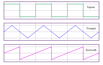
- Some PEMF therapy devices of sufficient intensity can induce or produce antimicrobial alternating currents (50-100 µA) in body tissue. E.g. SOTA® Magnetic Pulser or SOTA® Silver Pulser. These microcurrents can directly deactivate a large percentage of micro-organisms in the extra-cellular space. The maximum current is achieved when the EMF changes rapidly as in a square wave.
Leaves normal cells and beneficial enzymes intact and healthy. Human cells are very different from and much bigger than bacteria and viruses, and although the healthy cells do suffer transient disruption from the electromagnetic therapy, they are able to compensate for the disruption;
Promotes tissue repair / healing
Tissue injury impairs body’s healing biocurrents
- Weak bio-currents, naturally present in living organisms, control growth and repair. Healing bio-currents travel through the nerves or the liquid crystal semiconducting proteins of the connective tissue. Borgens et al, 1989; Ho, 1998; Oschman, 2000. Tissue injury or disease increases its electrical resistance, impeding the flow of these healing bio-currents through that tissue, and also decreases the transmembrane potential (“cell battery” voltage) of cells in damaged tissue Becker & Selden, 1985
- PEMF therapy produces or induces microcurrents in the body (of similar level to body’s endogenous healing currents) that have been shown to increase ATP production rate.
PEMF heals scar tissue
- Appropriate PEMF therapy can revert scar tissue to normal. A phenomenon of a repeatedly pulsed electromagnetic field is dedifferentiation of fibroblast cells and some types of precursor endothelial cell types into embryonic-looking cells. When a person suffers tissue damage, fibroblast cells migrate from body fatty tissue and the blood circulation to the damaged tissue site to form an emergency tissue patch. In minor damage, the injury will be nearly fully repaired with little scar tissue remaining in the area. Scar tissue mainly consists of fibroblast cells laid down to maintain a tough collagen protein fiber matrix, which holds the surrounding tissue together. However, when these cells are made to dedifferentiate by exposure to an appropriate PEMF, then during the time they are part of and maintaining scar tissue, or damaged tissue soon to become scar tissue, then some of the cells do not revert to being fibroblast and endothelial cells. Instead they become the type of cell that should be at the damaged tissue location or on the scar tissue edge, as if the damaged / scar tissue were not there.
- Repair time. By exposing the damaged tissue or scar tissue to repeated pulsed magnetic fields for ~5 to 15 minutes varying from twice every day to once every other day for approximately 1-6 weeks often much of the damaged tissue or scar tissue can be repaired /removed and be replaced with normal healthy tissue.
- A wide range of parameters are effective at reducing scar tissue. Experiments conducted by physicist Gary Wade at the Center for Complex Infectious Diseases showed that desired results could be obtained with a relatively wide range of combinations of range of pulsed electric field rise times, pulsed electric field strengths, electric field pulse time widths, pulse rates, and total time of exposure to pulses that could produce the desired results of having fibroblast cells become embryonic looking and embryonic-like in their behavior.
PEMF heals bone fractures / strengthens bones
- PEMF therapy is advocated for creating strong, healthy bones by stimulating osteoblasts to produce bone material.
Destroys cancer cells
Warning: Tumorous cancer cells must be killed off slowly. This gives the immune system time to deal with any dead microbes (and their former waste products) and the debris of destroyed cancer cells. Otherwise, the body can be dangerously overloaded with toxic waste.
PEMF therapy destroys existing cancer cells and prevents cells turning cancerous by deactivating microbes, increasing cell transmembrane “batteries”, and by “supercharging” body’s immune system. Note that most cells have a normal resting transmembrane potential (TMP or “cell battery” voltage) of ~70mV which is notably lower (by 15-20 mV) when a cell turns cancerous. It is interesting to note that heart cells maintain a TMP of ~100 mV, and do not turn cancerous.
Microbial hijacking of cells is a probable underlying cause of a cell turning cancerous. Microbial theory of cancer
Microcurrents cause cancer cells to become dehydrated. A prominent Swedish scientist, Bjorn Nordenstrom, found that cancer cells treated with electrotherapy appear to become dehydrated and die. He used an invasive surgical procedure, implanting electrodes in the tumor to generate microcurrents. (Note: PEMF therapy creates micro-currents in body tissue non-invasively). His basic techniques were adopted in China where, from 1988 to 1993 over 4,000 cases of advanced malignancy were treated. Of 2516 carcinoma cases in patients over 50 years old, a 78% favorable response rate was reported. Nordenstrom, 1987
Microcurrents alter cancer cell’s pH and DNA to deactivate reproduction. Dr. C.K. Chou, an American cancer researcher and his colleagues found that specific microcurrents alter the pH of the cancer cell and alter its DNA so that it cannot reproduce. Chou et al; 1997
Increases cellular pH (alkalizes body)
In order to function properly, your body necessarily keeps blood plasma pH in a narrow range pH range of 7.35 – 7.45.
TMP (Trans membrane potential or “Cell Battery” voltage) and alkalinity are connected. An alkaline environmment has a surplus of electrons. Conversely an acidic environment lacks electrons.
PEMF therapy normalizes the TMP, providing electrons to maintain a healthy pH in blood and body tissues. E.g. in so doing, the body does not need to rob bones and teeth of their strengthening alkalizing minerals.
Why is the body’s acid-alkaline balance of prime importance for health?
Other effects of PEMF therapy
Releases calcium ions from inside the cell. Calcium build-up inside artery wall cells contributes to plaque build-up in atherosclerosis. Adey, 1993
Pumping of lymph fluid by involuntary muscle contraction. Waste products of cellular metabolism and various proteins and mineral and elemental ions that need to be transported back into the general blood supply enter the lymph vessels from the extracellular space.
Regulates virtually every cell function
DNA, RNA and protein synthesis
Morphogenesis (concerned with shape of tissue, organs or organisms)
Stimulates hormonal production. PEMF therapy aids production of MELATONIN (by pineal gland) and HGH (Human Growth Hormone) by pituitary gland. Two essential hormones for sleep, a time when healing, regeneration and repair take place, all factors that affect energy levels and fight the aging process.
References
Adey WR. (1993, Apr.) Whispering Between Cells: Electromagnetic fields and regulatory mechanism in tissue. Frontier Perspectives; 3(2):21-25.
Adey WR., (1993 Apr) Biological effects of electromagnetic fields. J Cell Biochem. 51(4):410-6. Review.
Becker RO, Selden G. (1985) The Body Electric. New York: W. Morrow and Company Inc,
Borgens RB, Robinson KR, Vanable JW, McGinnis ME. (1989) Electric Fields in Vertebrate Repair. NY: Alan R. Liss
Bragin, Denis & L Statom, Gloria & Hagberg, Sean & Nemoto, Edwin. (2014). Increases in microvascular perfusion and tissue oxygenation via pulsed electromagnetic fields in the healthy rat brain. Journal of neurosurgery. 122. 1-9. 10.3171/2014.8.JNS132083. Pdf
Ceve G. (1990) “Membrane Electrostatics,”Biochim Biophys Acta, 103(3):311-82, Medline 91027827
Chou et al, 1997) Chou CK, McDougall JA, Ahn C, Vora N. Electrochemical treatment of mouse and rat fibrosarcomas with direct current. (1997) Bioelectromagnetics;18(1):14-24. PMID: 9125227.
CIESLAR G., SIERON A., TURCZYNSKI B., ADAMEK M. and JASKOLSKI F (1994 Nov). The influence of extremely low-frequency variable magnetic fields on rheologic and dielectric properties of blood and the water-electrolyte balance in experimental animals. Biochemistry and Bioenergetics. Vol 35, Issues 1-2. Pgs 29-32 Pdf
Culotta E & Koshland D Jr. (1992) NO news is good news. Science 258 (5090): 1862-1864 Link
Ho MW. (1998)The Rainbow and the Worm: The Physics of Organisms, 2nd ed. River Edge, NJ: World Scientific
Kirsch DL, PhD, DAAPM, FAIS (2011) Microcurrent Electrica Therapy Mechanisms and Results; Practical Pain Management. Link
Malzone A. et al, “Effect on cellular and tissue metabolism of induced electrical currents”Arch Stomatology 30(2):371-82 Medline 90314754
Ngok Cheng (1982) The Effect of Electric Currents on ATP Generation, Protein Synthesis and Membrane Transport in Rat Skin in Clinical Orthopedics, volume 171: pages 264-272. Link
Nordenstrom, Dr. Bjorn (1987) Electrochemical Treatment of Cancer. Link
Oschman JL. (2000) Energy Medicine: The Scientific Basis. Edinburgh, England: Churchill Livingstone.
Pan, Y. , Dong, Y. , Hou, W. , Ji, Z. , Zhi, K. , Yin, Z. , Wen, H. and Chen, Y. (2013), Effects of PEMF on microcirculation and angiogenesis in a model of acute hindlimb ischemia in diabetic rats. Bioelectromagnetics, 34: 180-188. doi:10.1002/bem.21755
Persinger MA (1974), ELF electric and magnetic field effects: ELF Electric and Magnetic Field Effects: The Patterns and the Problems. p. 275. Link
Yang M, Brackenbury, WJ (2013) Membrane potential and cancer progression. Front Physiol. 2013; 4: 185. pdf